What it was
This was a prototype Mask R-CNN system built to detect endotracheal tubes on neonatal chest X-rays and flag whether placement looked correct. It was one of the earliest clinical AI projects built as a proof of concept, before the difference between a model demo and a clinically safe workflow was fully understood.
What we built
The project used a Mask R-CNN style pipeline to localize the tube and produce image-level detections on chest radiographs. The report also includes the supporting architecture diagrams we used to reason about the workflow, including a vision-to-network concept figure and a Region Proposal Network diagram.
Model view
These figures show the technical side of the project: how the detection pipeline was framed and how the regions of interest were proposed before classification.
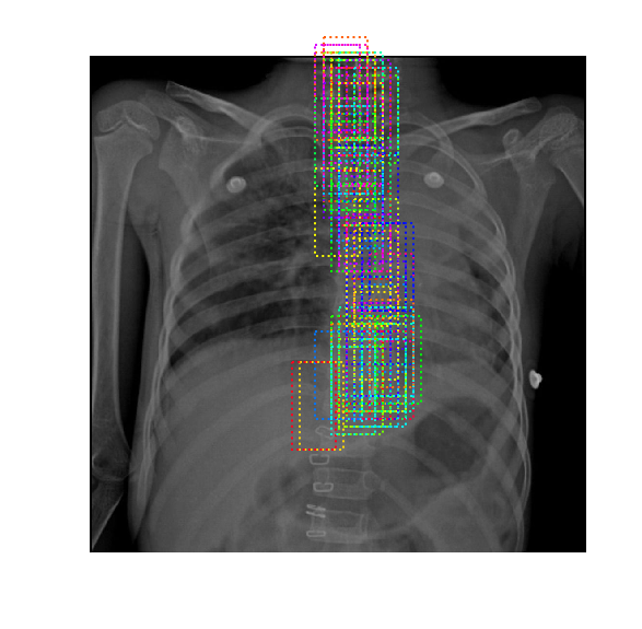
X-ray outputs
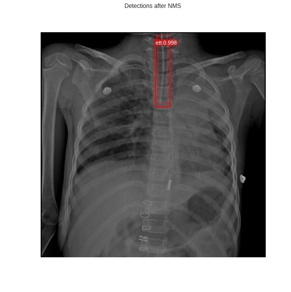
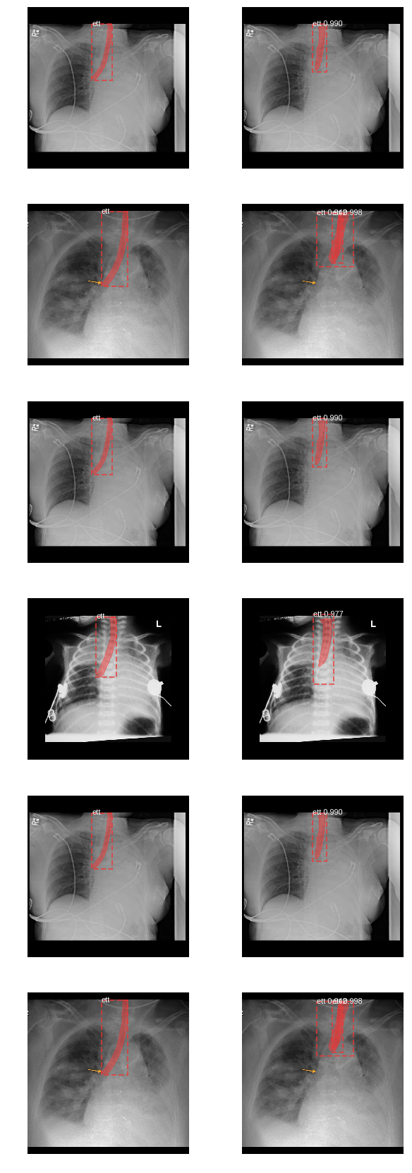
These images show the kind of radiographs the prototype was meant to inspect, including correctly placed tubes, model overlays, and harder cases with dense annotations.
Clean ET tube detection with a confidence label.
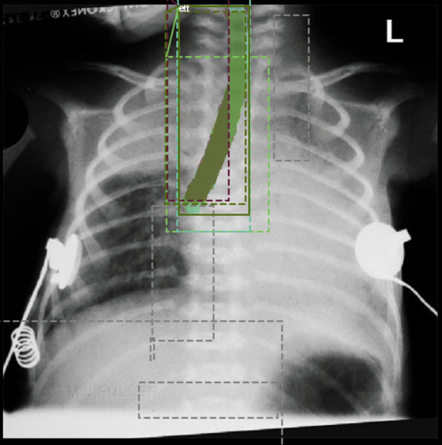
Chest X-ray with highlighted regions of interest.
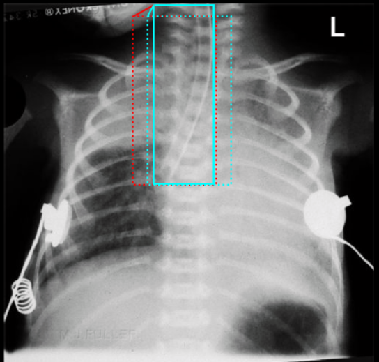
Grid of model outputs across multiple radiographs.
What it taught me
The main lesson was that clinical AI is not just about detection accuracy. The harder problems are consistency, workflow fit, and deciding when a model is good enough to be trusted in a high-stakes setting.
Why it is deprecated
This project is now presented as a historical prototype rather than an active tool. It reflects an early stage in the work: useful for learning, important to show, but not something to position as production-ready clinical software.